Fetal Cardiology Assessment


As part of my role as a paediatric and fetal cardiologist I see pregnant women in our fetal cardiology clinics in GOSH and the combined fetal medicine and fetal cardiology clinic at UCLH.

The advancement in scanning technology means that many congenital heart problems or fetal heart rate and rhythm issues, are picked up before the baby is born.


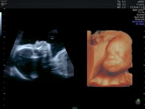
NORMAL FETAL HEART AT 20 WEEKS

Normal views of Aortic arch in 20 week fetus


In some cases we screen mothers in whom there is a known risk factor that puts their baby at an increased risk of a congenital heart defect, these include a family history of congenital heart disease, certain maternal diseases such as diabetes or certain connective tissue diseases. In addition findings on routine scanning and combined screening testing early in pregnancy may suggest an increased risk of congenital heart disease.

I understand it can be very worrying and distressing to be told there may be something wrong with your baby’s heart. Fetal cardiology provides expert evaluation and diagnosis of fetal heart defects. With explanation of and counseling about all treatment options. It then allows careful planning for your baby’s delivery. The parents can avail of a range of support services to help support the family before and after the baby’s delivery.

The optimal timing for fetal echocardiography is 18-20 weeks gestation. Sometimes if the fetus is in a good position, adequate images can be obtained earlier.

Normal fetal echocardiogram at 16 weeks gestation
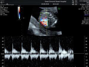
Fetal echocardiogram at 16 weeks demonstrating normal fetal heart rate and rhythm


NORMAL 4 CHAMBER HEART AT 16 WEEKS


NORMAL AORTIC ARCH 16 WEEKS
Congenital Heart Disease

Congenital heart disease describes a range of birth defects that affect the normal workings of the heart. It can affect 8 in every 1000 babies born. Symptoms are noticeable soon after birth, although mild defects may not cause any problems until later in life. If there are concerning symptoms a referral is usually made to a paediatric cardiologist for further evaluation.

What causes congenital heart disease?

The exact cause of congenital heart disease in children is often not found and is likely to be multifactorial. These may include a genetic basis, environmental factors or infection. A positive family history of heart problems in children, or symptoms and signs such as a murmur, cyanosis or breathlessness are often common reason to seek review.

Most paediatric cardiology problems in children are congenital in nature (something people are born with) due to a problem in the way the heart was put together during the very early stages of pregnancy. If you have a child with a heart problem or concerns regarding a possible cardiac issue and are considering pursuing private care, I welcome any enquiries and I can be contacted by email, or via my practice manager.

Is it always awful news?

Fortunately, the majority of complaints and symptoms observed in children are infrequently associated with any underlying congenital cardiac condition. A few simple tests performed by a paediatric cardiologist (a child heart specialist doctor) are able to exclude all significant congenital cardiac conditions.

Echocardiography
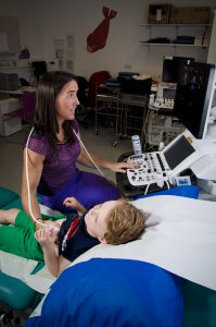
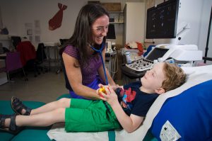
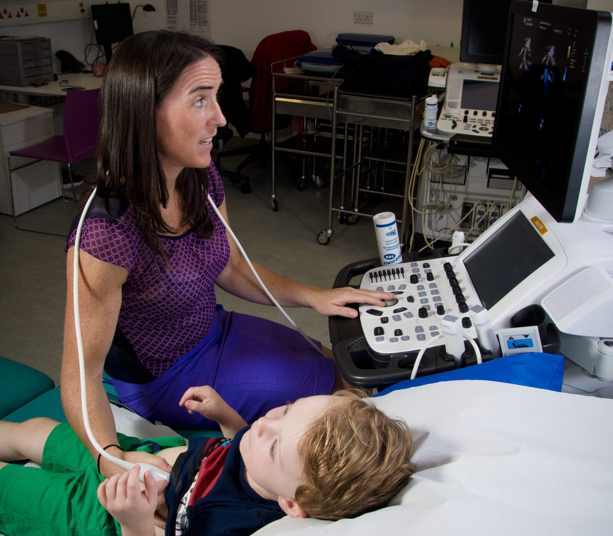
Paediatric Echocardiography By Dr Michelle Carr

Heart Murmur

What is a heart murmur?
A heart murmur is a sound made by blood moving through the heart’s chambers or valves. Murmurs range from quite faint to very loud.

There are two main types of heart murmurs: innocent or flow (harmless) and abnormal. Most commonly heart murmurs in children are innocent heart murmurs that are not caused by a heart problem. They tend to become less pronounced and are less frequently as the child gets bigger.

Most abnormal murmurs in children are due to a congenital heart defect or a vascular anomaly.

What causes a heart murmur?
Heart murmurs may be caused by a number of conditions related to turbulent accelerated flow of blood in the heart, some examples include:

heart valve problems
holes between the pumping chambers
narrowing of blood vessels leaving the heart
peripheral pulmonary stenosis
systemic disease such as: fever or anaemia

How is a heart murmur diagnosed?
A physician will evaluate your child’s murmur based on what it sounds like  (pitch, character, location, loudness and duration). Most are detected in the first instance by a primary care doctor. Your GP or paediatrician will decide if your child needs to be seen by a cardiologist. The most important thing early on is to diagnose the cause of the murmur.

Murmurs related to a congenital heart defect or other problem involving the heart will be heard the loudest in the area of the chest where the problem occurs.

What are the treatment options for a heart murmur?
Luckily, most heart murmurs in children are benign and don’t need treatment or follow up. If your child has an abnormal heart murmur that’s caused by another heart condition, he or she may need further investigations to fully define the cause and assess the necessary treatment for that condition.

Treatment of abnormal heart murmurs?
If your child has a heart murmur caused by a heart condition, the necessary investigations will be performed to allow counselling and appropriate care for the child depending on the underlying lesion causing the murmur.

Echocardiography
Paediatric Echocardiography By Dr Michelle Carr

Ventricular Septal Defect (VSD) – “hole in the heart”

What is a ventricular septal defect?
A ventricular septal defect (VSD) is a hole in the wall that separates the lower right and left heart pumping chambers (ventricles). It is the most common type of congenital (present from birth) heart condition.

In patients with VSD, oxygen-rich blood passes from the left ventricle and mixes with oxygen-poor blood in the right ventricle. This sends the blood back to the lungs and makes the work of the heart inefficient and harder. The larger the hole, the more symptoms it can cause. If there is significant flow of blood across the hole  some infants may develop difficulty with growth and breathing. Symptoms due to the VSDs may be able to be managed with medication and high calorie feeding regimes. If that is not sufficient, surgical repair to close the hole is recommended. Rarely, a VSD can lead to an infection in the heart, called bacterial endocarditis.

Treatment for babies and children with ventricular septal defects?
Many VSDs are small enough that observation or medical therapy, including higher calorie formula or medications to relieve congested breathing, are all that is needed.  Fro many VSDs the natiral history is for them to become smaller on their own and even close spontaneously over time.

If an infant has significant difficulties with growth or breathing, despite medical therapy, then surgical closure can be performed with excellent results.  In some instances the defect can be amenabke to a catheter or “key-hole” based procedure. The need for surgical or interventional closure is based on the type and size of defect as well as clinical symptoms seen in the baby or child.

Atrial Septal Defect (ASD)

What is an atrial septal defect?
An atrial septal defect (ASD) is a hole in the wall that separates the upper right and left heart collecting chambers (atria). “Atrial” is the name we give to the upper chambers of the heart, “septal” is a medical term for wall, and “defect” is a gap. Taken together, it means that there is a gap in the wall of the upper portion of the heart.

ASD is a congenital condition present at birth. The muscle wall dividing the two upper chambers (atria) has multiple building blocks and a deficiency in any of them can lead to a gap in a different portion of this partitioning upper wall.

The ASD allows oxygen-rich (red) blood to pass from the left heart chamber and mix with oxygen-poor (blue) blood in the right chamber.  The effects this communication has on the child’s heart depends on the size of the hole. Some communications are too small to of any significance, the most common type we see is called a PFO (patent foramen ovale). This can be found in up to 1 in 3 adults and is considered a normal variant. If the communication allows a significant amount of blood cross from the left heart to the right heart over time the right heart will increase in size and work harder. Generally this is tolerated quite well initially but if left over time, this can cause the heart and lungs to work harder and put the child at risk for other conditions later in life,  as an adult. These include:

abnormal cardiac rhythms
heart muscle failure
reduced exercise tolerance
pulmonary hypertension

Treatment of ASDs?
As mentioned, the size of the hole can vary from small to large. Some ASDs close on their own over time, typically during the first three years of life. However, if the hole remains significant in size, closure is necessary. The options include non-surgical, catheter based “key-hole” interventions and minimally-invasive surgical techniques. Depending on the size and location of the ASD, non-surgical catheter-based techniques may be able to close the defect, thereby avoiding surgery. Each ASD is assessed individually to ascertain the most appropriate management and closure option.


AVSD: atrioventricular septal defect

An atrioventricular septal defect or AVSD is a combination of several associated lesions that are affected in the development of the valves between the top chambers (atria) and bottom chambers (ventricles) that result in a large defect in the centre of the heart. Also known as an endocardial cushion defect, the condition is congenital, which means it is present at birth, and occurs in two out of every 10,000 newborns. There is a strong association between this defect and the chromosomal abnormality, Down Syndrome.
When the heart is properly septated, red oxygenated blood from the lungs does not mix with the blue deoxygenated blood from the body. In an AVSD blood can move and mix freely among the four heart chambers. If left untreated, AVSD can cause inefficient blood flow through the heart and ultimately to

AVSD is not a single defect but rather a group of closely-associated defects in various combinations and with varying degrees of severity:
-Atrial component (communication between the top chambers)
-Ventricular component (communication between the lower chambers)
-AV valve (mitral and tricuspid) abnormality, failure of normal separation into 2 distinct particioed valves that separate the upper heart chambers (atria) from the lower chambers (ventricles), often resulting in one large “common” valve sitting over both right and left chambers.

How do we treat AVSDs
All of the AVSD subtypes, usually require surgery. The surgery, involves closure of any holes in the chambers, using patches. If the left sided AV valve does not close completely, it is repaired to make it more competent. The surgeon repairs the the common valve, separating it into two distinct valves – one on the right side and one on the left side.

The age at which surgery is dependent on clinical factors such as the child’s health and growth as well as the specific structure of the AVSD.  Ideally, surgery should be done before there is permanent damage to the lungs from too much blood being pumped to the lungs. Medication may be used to treat congestive heart failure, but it is only a short term measure in an attempt to allow the infant become bigger and strong enough for surgery.

Infants who have surgical repairs for AVSD generally remain under lifelong follow up. Some of the defects can be more complicated and there is a discrepancy in size between the 2 valves and the lower chambers, this is termed an unbalanced AVSD and can be more challenging to treat. Each AVSD is individual and the medical and surgical management are tailored to the defect. In some instances, children can require more than one operation. The most common need for reintervention is for complications of a leaky left AV valve. A child or adult with an AVSD will need regular follow-up visits with a cardiologist to monitor his or her progress, avoid complications, and check for other health conditions that might develop as the child gets older. With proper treatment, most babies with AVSD grow up to lead healthy, productive lives.

Coarctation

What is coarctation of the aorta?
The aorta is the major blood vessel carrying oxygenated (red) blood to the body. Coarctation of the aorta refers to a condition where there is a tightness (or narrowing) in this body artery. A coarctation is usually located just past the aortic arch, which has branches providing blood to the head and arms. When this defect is present, blood flow is restricted to the lower half of the body and the left ventricle muscle of the heart must pump harder to force the blood through the narrowed opening.

The clinical presentation is variable and depends principally upon the severity of the narrowing and the age of the baby or child. Milder degrees of coarctation are often not detected until childhood or adolescence and often come to light from the incidental finding ofweaker leg (femoral) pulses, a murmur or hypertension (high blood pressure). When the condition is severe, there can be reduced blood flow to the body in these children. In babies, this blockage can cause the left ventricle to weaken and make your baby very sick. In older children, this blockage can cause the left ventricle to thicken over time and also cause high blood pressure in the brain. If the muscle becomes too thick and is no longer able to function well, it can eventually fail.

Coarctation of the aorta occurs in about 6 to 8 percent of all children with congenital heart disease, and twice as frequently in boys. It also occurs in about 10 percent of girls who have Turner syndrome, a chromosomal abnormality.

Seventy-five percent of children with coarctation of the aorta also have a bicuspid aortic valve, in which the aortic valve has two leaflets, instead of the usual three. It can also be associated with other left sided heart defects and ventricular septal defects.

Treatment for coarctation of the aorta?
The majority of coarctations are treated surgically, although in older children who are suitable,  there is increasing experience with catheter-based (key-hole) interventions as well. Treatment strategies are tailored to the child’s age, size and the anatomy of the aortic arch as well as the presence of other associated cardiac conditions.

Patent Ductus Arteriosus (PDA)

What is patent ductus arteriosus?
Patent ductus arteriosus is a congenital heart condition where there is a persistent connection between the pulmonary artery and the aorta. This causes blood to mix between the two arteries and forces the heart and lungs to work harder. This special tube is necessary in fetal life before baby’s are born to allow them to use the placenta.  Usually, it is programmed to close spontaneously in the first few days of life after birth.

Patent ductus is the sixth most common congenital heart defect. It occurs in 5 to 10 percent of all children born with congenital heart disease. Patent ductus occurs twice as often in girls as in boys. It is more common in premature infants.

In babies with patent ductus, the fetal ductus arteriosus remains open (patent). This allows the oxygen-rich (red) blood to mix with oxygen-poor (blue) blood and forces the lungs and heart to handle a larger amount of blood than is normal. This is inefficient flow and causes the heart to work harder to meet it’s demands. If the PDA is large and if left over time it  can lead to heart failure.


Treatment children with a patent ductus arteriosus?
If the PDA is large enough to cause a haemodynamic burden on the child’s heart then we would advise to close the communication. Treatments, including medical therapy, minimally-invasive catheter-based interventions and minimally-invasive surgical solutions are available depending on the size and type of PDA.

Chest pain

Although the symptom of chest pain in your child can set off alarm bells in most parents, studies have shown in contrast to adults, the overwhelming majority of cases of chest pain in otherwise healthy children have a noncardiac aetiology. Numerous previous studies in multiple practice settings have demonstrated a cardiac aetiology in only 0-5% of children and adolescents presenting with chest pain.

True cardiac chest pain is rare in a teenager or child. A detailed clinical history of the nature, type and frequency of the pain can often help a padiatric cardiologist stratify whether the pain has an underlying cardiac cause. In the vast majority, a careful focused history and physical examination, along with a screening tests such as an ECG, will identify essentially all patients with a serious cardiac condition.

A 12 lead ECG (or electrocardiogram) is a valuable initial investiugation. This test involves placing 12 stickers or electrodes on the chest and analysing how the electrical impulses spread through the heart. The presence of other symptoms such as palpitations (an irregular or fast heart rate) or a history of syncope or collapse would prompt more detailed investigation and assessment. A positive family history of cardiac problems or history of early / unexplained death would also be a reason for further investigations.

Arrhythmias in Children

What is an arrhythmia?
An arrhythmia is an abnormal rhythm of the heart, which can cause the heart to pump less effectively. It most often occurs in adults but can also occur in children. Some arrhythmias are not dangerous, while others can be life threatening.

Basic ECG

Arrhythmias are classified by the area of the heart where they start: the upper chambers (atria) or lower chambers (ventricles). They can be related to the heart beating too fast (tachycardia), too slow (bradycardia) or in an irregular pattern (fibrillation).

There are many types of arrhythmias, including:

atrial flutter
atrial fibrillation
sinus arrhythmia
sinus tachycardia
sick sinus syndrome
premature supraventricular contractions or premature atrial contractions (PAC)
supraventricular tachycardia (SVT) or paroxysmal atrial tachycardia (PAT)
premature ventricular contractions
ventricular tachycardia
ventricular fibrillation


What are the symptoms of an arrhythmia?

Some arrhythmias cause no symptoms. When they do, symptoms can include:

fatigue
rapid breathing
palpitations
dizziness
fainting

What are the causes of an arrhythmia?
Arrhythmias occur when there is a problem with the electrical signals that control the heartbeat. Sometimes the nerve cells that create the electrical signals don’t work the way they should or don’t move normally through the heart. In some cases, other areas of the heart begin to produce electrical signals that disrupt the normal heartbeat.

There is a specific Electrophysiology team with a special interest in complex paediatric rhythm issues. If necessary a referral can be made to one of this team to further investigate and treat these conditions.